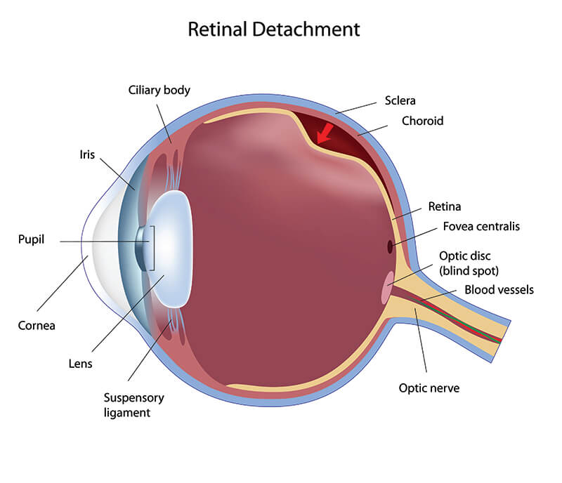
The retina is the light-sensitive layer of tissue that lines the inside of your eye. It is composed of nerve fibers that are responsible for transmitting nerve impulses to the optic nerve so that they can be converted to visual messages in the brain. Under normal circumstances, the retina is attached to underlying tissue so that it “lines” the inner surface of the eye. If the retina is lifted or pulled away from the underlying tissue, it is called a Retinal Detachment.

The Prevalence and Risk of Retinal Detachment
In normal, healthy eyes, the risk of Retinal Detachment is about 5 per 100,000 per year with a greater frequency in the middle-aged or elderly population of perhaps 20 per 100,000 per year. Retinal Detachment is more frequent if you are myopic or nearsighted and especially if your prescription is above 6.00 Diopters of correction. In fact, 67% of Retinal Detachment cases occur in myopic eyes.
Further, Retinal Detachment is more prone to occur in association with certain eye conditions and diseases including after cataract surgery and in patients suffering from Diabetic Retinopathy. Fortunately, with modern cataract surgery and early treatment of Diabetic Retinopathy, the incidence of Retinal Detachment is low.
Retinal Detachment Symptoms
Retinal detachment is a painless sight-threatening eye problem that causes a number of symptoms and warning signs that occur often before the actual detachment happens. The key to preserving vision is to recognize these warning signs to schedule an exam for quick diagnosis and treatment. Warning signs and symptoms may include:


- Flashes of light that may occur in your field of vision toward the outermost periphery.
- A vitreous detachment may appear as a sudden increase in the number of floaters in your vision and possibly even a ring of floaters or “hairs” in your vision-sometimes this is accompanied by “specks” or a “cobweb”.
- A retinal detachment may appear as the sense of a “shadow” in your peripheral vision that may progress toward the center of your vision or a sensation of a “curtain” or a “veil” being drawn over your vision.
- In extreme cases of retinal detachment, you may experience a loss of central vision.
Types of Retinal Detachment
The Vitreoretinal Surgeons at Eyecare Medical Group in Portland typically see three types of retinal detachment. These include:
- Rhegmatogenous Retinal Detachment
- Exudative, Serous or Secondary Retinal Detachment
- Tractional Retinal Detachment
Rhegmatogenous Retinal Detachment occurs as a result of a break (usually a tear or hole) in the Retina that permits fluid to pass into the space underneath the Retina. Tears or holes in the Retina may actually occur without causing any symptoms to occur. Therefore, it is important that you have routine eye examinations, especially if you are nearsighted or myopic, or if you play contact sports and might be subjected to eye trauma, in order to rule out the presence of Retinal Breaks before they cause loss of vision. Furthermore, if you are nearsighted or myopic, you may be more prone to Peripheral Retinal Degenerations, of which Lattice Degeneration may also increase your risk of Retinal Detachment making regular eye examinations an even more important part of your routine health care. Rhegmatogenous Retinal Detachment is the most common type of Retinal Detachment.
Exudative Retinal Detachment may occur due to inflammation, injury or a Retinal Vascular Disease that causes fluid accumulation underneath the Retina without the presence of a Retinal Hole or Retinal Tear.
Tractional Retinal Detachment may occur when fibrous or fibrovascular scar tissue has been formed on the Retina as a result of an injury, inflammatory disease or the presence of neovascularization, such as in Diabetic Retinopathy. The scar tissue actually pulls the Retina free from the underlying pigment layer it is normally attached to, causing a Retinal Detachment.
Treatment of Retinal Detachment
The Vitreoretinal Surgeons at Eyecare Medical Group in Portland have extensive experience in all types of treatment for retinal detachment which they can perform in the Outpatient Eye Surgery Center for patients from throughout Maine, Vermont, New Hampshire, and northern New England. Depending on the cause and the location as well as the degree of detachment, our Retina Specialists might recommend:
- Cryopexy and Laser Retinopexy are often used to create an adhesion or scar around the edge of a Retinal Hole, or small Retinal Tear in order to prevent fluid from passing through the area and underneath the Retina and causing a Retinal Detachment. Cryopexy uses a “cold” instrument, about the size of a pencil, to freeze the damaged area whereas Laser Retinopexy uses a powerful “laser beam” to achieve the creation of the tiny scar. Generally, these two treatments are interchangeable but Cryopexy is often used if there is a considerable accumulation of fluid.
- Pneumatic Retinopexy is another type of retinal surgery for repairing Retinal Detachment. During Pneumatic Retinopexy, the Retina Surgeon injects a “gas bubble’ into the back of the eye after first applying the Laser or Cold treatment. The patient’s head is positioned so that the “gas bubble” acts to place gentle pressure against the damaged area, allowing the fluid underneath to be absorbed and the Retina to reattach. This often requires that patients lie perfectly still in a particular position for several days or even a week in order for the results to take effect.
- Scleral Buckle Surgery is a well-established treatment for Retinal Detachment whereby the Retinal Surgeon places a silicone band around the eye to gently apply pressure to the outermost walls of the eye, allowing the retinal breaks to close and the Retina to reattach. Scleral Buckle Surgery may be done in conjunction with Cryopexy or Laser Photocoagulation to assure the best possible results.
- Vitrectomy is an increasingly common treatment for Retinal Detachment that involves surgically removing the Vitreous gel in the back part of the eye in conjunction with the injection of a “gas bubble.” Vitrectomy surgery can lead to the formation of a Cataract.
NOTE ABOUT RETINAL DETACHMENT TREATMENT
After surgery for Retinal Detachment, the results can often take many weeks for patients to achieve their visual recovery. In certain cases, the vision after Retinal Detachment Surgery may not allow the full recovery of precise vision, especially if the Macula was detached in addition to other areas of the Retina. Early diagnosis and rapid treatment are the keys to preventing vision loss from Retinal Detachment. Regular eye examinations are an excellent way to learn if you are at risk for Retinal Detachment.


